Guide Navigation
- 1. An Introduction to the Hematocrit Centrifuge
- 2. The Scientific Principle: How a Hematocrit Centrifuge Separates Blood
- 3. Anatomy of a Hematocrit Centrifuge: Components and Technical Specifications
- 4. The Microhematocrit Method: A Step-by-Step Procedural Guide
- 5. Applications in Clinical Diagnostics and Research
- 6. How to Select a Hematocrit Centrifuge for Your Laboratory
- 7. Essential Maintenance, Calibration, and Safety Protocols
- 8. Conclusion: A Fundamental Tool for Accurate Blood Analysis
 |
A hematocrit centrifuge (Browse HINOTEK advanced Hematocrit Centrifuge) is a core piece of equipment in hematology and clinical diagnostics. Its function is specific and critical: to determine the volume percentage of red blood cells within a whole blood sample. This value, known as the hematocrit or Packed Cell Volume (PCV), is a fundamental indicator of a person’s health. This page provides a comprehensive overview of the hematocrit centrifuge, covering its operating principles, components, applications, and the standard procedures for its use in a laboratory setting.
An Introduction to the Hematocrit Centrifuge
What is a Hematocrit Centrifuge?
A hematocrit centrifuge is a specialized laboratory instrument designed to separate whole blood into its constituent components by spinning it at high speeds. Unlike general-purpose centrifuges that accommodate a variety of tube sizes and applications, the hematocrit centrifuge is optimized for one primary task. It uses small-bore glass or plastic capillary tubes to hold blood samples and applies high centrifugal force to achieve a rapid and precise separation. The result is a clear measurement of the packed cell volume (PCV), which is the proportion of the total blood volume occupied by red blood cells. This specialization ensures high accuracy and reproducibility for one of the most common blood tests performed in clinical settings.
Understanding Hematocrit (Packed Cell Volume)
Whole blood is a complex fluid composed of several key components: red blood cells (erythrocytes), white blood cells (leukocytes), platelets (thrombocytes), and the liquid medium in which they are suspended, called plasma. The hematocrit value specifically measures the volume of red blood cells, expressed as a percentage of the total volume.
Red blood cells contain hemoglobin, the protein responsible for transporting oxygen from the lungs to the rest of the body. The volume of these cells is therefore a direct indicator of the blood’s oxygen-carrying capacity. An abnormal hematocrit level can signal various medical conditions, making its measurement a crucial first step in many diagnostic pathways.
The Role in Modern Laboratory Diagnostics
The hematocrit centrifuge is a fundamental tool in a wide range of settings, including clinical laboratories, hospitals, blood banks, and research facilities. While modern laboratories often use large automated hematology analyzers to calculate hematocrit based on red blood cell count and volume, the manual microhematocrit method performed with a centrifuge remains indispensable.
It serves as a reliable backup method, a gold standard for calibrating automated analyzers, and the primary testing method in smaller clinics, field laboratories, or regions with limited access to automated equipment. The test’s speed is a significant advantage; a precise hematocrit value can be obtained in minutes, providing physicians with critical information for immediate patient care and treatment decisions.
The Scientific Principle: How a Hematocrit Centrifuge Separates Blood
The operation of a hematocrit centrifuge is based on a straightforward physical principle: sedimentation under centrifugal force. By spinning blood samples at a high velocity, the instrument generates a force many thousands of times greater than gravity, causing the components of the blood to separate according to their density.
The Physics of Centrifugal Force
When the centrifuge rotor spins, it subjects the capillary tubes to a strong centrifugal force, which acts outwards from the center of rotation. This force compels denser particles to migrate further away from the center, while less dense components remain closer to it. In a blood sample, the cellular components (red cells, white cells, platelets) are denser than the liquid plasma. The centrifuge exploits these small differences in density to achieve a clean separation.
The Three Layers of Centrifuged Blood
After centrifugation is complete, the blood within the capillary tube visibly separates into three distinct layers. Observing these layers provides both quantitative data and qualitative diagnostic clues.
- Layer 1: Packed Red Blood Cells (Erythrocytes)At the bottom of the tube is the densest layer, a column of tightly packed red blood cells. This layer is dark red and represents the packed cell volume, which is the component that is measured to determine the hematocrit.
- Layer 2: The Buffy CoatAbove the red blood cells is a very thin, grayish-white middle layer known as the buffy coat. This layer contains the white blood cells and platelets, which are less dense than red blood cells but denser than plasma. While the buffy coat is not included in the hematocrit measurement, its appearance is clinically significant. A visibly thickened buffy coat can indicate a high white blood cell count (leukocytosis), which may suggest an infection or a condition like leukemia.
- Layer 3: PlasmaThe top layer is the plasma, the least dense component of blood. It is a clear, straw-colored liquid that consists of water, proteins, electrolytes, and other solutes. The color of the plasma can also provide diagnostic information; for example, a reddish tint may indicate hemolysis (ruptured red blood cells), while a cloudy appearance may suggest high lipid levels (lipemia).
Calculating the Hematocrit Value
The hematocrit value is calculated as a ratio of the packed red cell volume to the total volume of the sample. The procedure is as follows: the height of the packed red blood cell column is measured and then divided by the total height of the blood column, which includes the red cells, buffy coat, and plasma. This ratio is then multiplied by 100 to be expressed as a percentage.
The formula is:
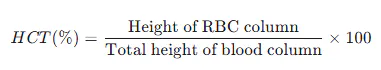 |
To ensure accuracy, this measurement is not done by eye. It is performed using a specialized microhematocrit reader card or a built-in digital reader, which allows for precise alignment and calculation. It is critical to exclude the buffy coat from the red blood cell measurement, as its inclusion is a common source of falsely elevated results.
Anatomy of a Hematocrit Centrifuge: Components and Technical Specifications
A hematocrit centrifuge is engineered for a specific purpose, and its components reflect this focus on precision, safety, and efficiency. Understanding the function of each part and its key technical specifications is essential for operators and procurement staff.
Core Components and Their Functions
-
- Motor: Modern hematocrit centrifuges are equipped with a brushless DC motor. This type of motor is preferred for its long operational life, minimal maintenance requirements, precise speed control, and quiet operation, which contributes to a better laboratory environment.
 |
- Rotor: The rotor is the most specialized component. It is a fixed-angle rotor designed to hold a specific number of capillary tubes, typically 24. A key design feature is the inclusion of individual chambers or support trays for each tube. This thoughtful design contains any glass fragments or blood leakage in the event a tube breaks, preventing a chain reaction that could damage other samples and contaminate the centrifuge. During loading, the sealed ends of the capillary tubes must always face the outer perimeter of the rotor.
- Control Panel & Display: The user interface typically features a large, clear LCD screen and intuitive controls. This allows the operator to easily set and monitor essential parameters such as speed in Revolutions Per Minute (RPM), Relative Centrifugal Force (RCF), and run time. Many models allow the user to toggle the display between RPM and RCF during a run, providing real-time operational data.
- Lid and Safety Lock: A robust lid with an electronic or mechanical safety interlock is a critical safety feature. This mechanism prevents the centrifuge from starting if the lid is not properly closed and ensures the lid cannot be opened while the rotor is in motion. On many models, the lid unlocks and opens automatically once the run is complete and the rotor has stopped.
- Housing: The centrifuge body is typically constructed from sturdy steel or a durable plastic-metal composite. The design prioritizes a small footprint to conserve valuable laboratory bench space.
Understanding Key Technical Specifications
When evaluating a hematocrit centrifuge, several technical specifications are paramount. The most critical parameter for achieving proper separation is RCF, not RPM. RCF accounts for the radius of the rotor, providing a standardized measure of the force applied to the sample. A protocol specifying an RCF is more accurate and reproducible than one specifying only an RPM, as two different centrifuges running at the same RPM may generate different RCFs if their rotor sizes differ.
- Speed (RPM): This measures how fast the rotor spins in Revolutions Per Minute. Hematocrit centrifuges are high-speed devices, with typical ranges from 500 RPM up to 12,000 RPM or higher.
- Relative Centrifugal Force (RCF or x g): This is the actual force exerted on the samples, measured in multiples of Earth’s gravitational force (g). For complete packing of red blood cells, an RCF between 11,000 and 15,000 x g is typically required.
- Capacity: This refers to the number of capillary tubes the rotor can process in a single run. The standard capacity is 24 tubes, which allows for efficient batch processing and high throughput in busy labs.
- Timer: A digital timer allows the operator to set the precise duration of the centrifugation cycle. Run times are typically short, with timers ranging from 0 up to 30 minutes, though a standard spin is only 4 to 5 minutes.
- Safety Features: Beyond the essential lid lock, modern centrifuges include imbalance detection, which automatically powers down the unit if an unbalanced load is detected, preventing mechanical damage and ensuring operator safety.
Table 1: Typical Technical Specifications of a Hematocrit Centrifuge
This table summarizes the key specifications for a typical hematocrit centrifuge, providing a quick reference for comparison and purchasing decisions.
| Feature | Typical Range/Value | Importance in the Lab |
| Motor Type | Brushless DC | Low maintenance, quiet operation, long lifespan, precise speed control. |
| Max Speed (RPM) | 12,000 – 14,000 RPM | Provides the high velocity needed to generate sufficient separation force. |
| Max RCF (x g) | 12,000 – 15,300 x g | The critical measure of force required for complete and rapid packing of red blood cells. |
| Rotor Capacity | 24 Capillary Tubes | Determines sample throughput; a 24-place rotor is standard for clinical efficiency. |
| Timer Range | 0 – 30 minutes | Allows for precise, repeatable run times, ensuring consistent results. |
| Key Safety Features | Imbalance Detection, Lid Lock | Protects the user and the instrument from damage due to operational errors. |
The Microhematocrit Method: A Step-by-Step Procedural Guide
Performing the microhematocrit test requires careful attention to detail at every step to ensure an accurate result. The most common sources of error in this procedure are pre-analytical, meaning they occur before the sample is even placed in the centrifuge. Following a strict standard operating procedure (SOP) is the best way to minimize these errors.
Required Materials
Before beginning the procedure, gather all necessary materials :
- Hematocrit centrifuge
- Microhematocrit capillary tubes (heparinized or plain)
- Sealing clay or compound
- Microhematocrit reader (card or digital)
- Personal Protective Equipment (PPE), including gloves
- Gauze or lab wipes
- Sharps container for disposal
Step 1: Sample Collection and Tube Selection
The first critical decision is selecting the correct capillary tube based on the blood sample source. This choice prevents errors related to blood clotting or sample dilution.
- Sample Source: Blood can be collected via venipuncture into a tube containing an anticoagulant like EDTA (identified by a purple top) or directly from a skin puncture (a finger or heel stick).
- Heparinized Tubes (Red Tip): These tubes are internally coated with the anticoagulant heparin. They must be used when collecting blood directly from a skin puncture. Without the heparin, the fresh blood sample would clot inside the narrow tube before it could be centrifuged.
- Non-Heparinized/Plain Tubes (Blue Tip): These tubes have no anticoagulant coating. They must be used when the blood sample is taken from a pre-anticoagulated source, such as an EDTA tube. Using a heparinized tube with an already anticoagulated sample would introduce excess anticoagulant, potentially diluting the sample and causing a falsely low hematocrit reading.
Step 2: Filling and Sealing the Capillary Tube
Proper filling and sealing are essential to prevent sample loss during centrifugation.
- Mix the Sample: If using blood from an EDTA tube, gently invert the tube 8-10 times to ensure the cells are evenly suspended.
- Fill the Tube: Hold the capillary tube horizontally with a slight downward tilt. Touch the open end (the end without the colored ring) to the drop of blood. Capillary action will draw the blood into the tube. Allow it to fill to approximately two-thirds or three-quarters of its length. Avoid introducing any air bubbles, as they can interfere with the reading.
- Seal the Tube: Wipe any excess blood from the outside of the tube with a clean gauze pad. Seal the dry end of the tube (the end that was not in the blood) by pressing it vertically into a tray of sealing clay. The plug should be at least 4 mm long to create a secure seal and prevent leakage.
Step 3: Loading and Balancing the Centrifuge
Balancing the rotor is the single most important step for safe centrifuge operation. An unbalanced load can cause severe vibration, damage the instrument, and lead to sample breakage.
- Place Tubes Symmetrically: Place the filled capillary tubes into the rotor slots. Each tube must be counterbalanced by another tube placed in the diametrically opposite slot.
- Use a Balance Tube: If you have an odd number of samples, you must create and use a “balance tube.” This is a spare capillary tube filled with water or blood to the same level as your sample tube.
- Correct Orientation: Place the tubes in the rotor with the sealed (clay) end facing outwards, against the rubber gasket at the perimeter of the rotor. This ensures the force pushes the blood column against the seal.
- Secure the Lid: Place the inner rotor lid on top and tighten it securely. Then, close and lock the main centrifuge lid.
Step 4: Centrifugation
- Set Parameters: Set the centrifuge to the required speed and time. A typical setting is a force of 10,000 to 15,000 x g (which corresponds to roughly 11,000 to 12,000 RPM on most models) for a duration of 4 to 5 minutes. These parameters are designed to achieve maximum packing of the red blood cells.
- Start the Run: Press the start button to begin centrifugation.
- Wait for Completion: Do not attempt to open the lid while the centrifuge is running. Wait until the cycle is complete and the rotor has come to a full stop.
Step 5: Reading the Hematocrit Result
Immediately after centrifugation, the tubes should be read to prevent the packed cells from resettling.
- Position the Tube: Carefully remove a capillary tube from the rotor and place it on the microhematocrit reader card or in the groove of a digital reader.
- Align the 0% Line: Adjust the tube so that the bottom of the packed red blood cell column (the interface between the red cells and the sealing clay) aligns perfectly with the 0% line on the scale.
- Align the 100% Line: Adjust the scale or a movable line on the reader so that the top of the plasma column (the meniscus) aligns with the 100% line.
- Read the Result: The hematocrit value is read directly from the scale at the point corresponding to the top of the packed red blood cell layer, just below the buffy coat.
- Check for Agreement: The procedure should be performed in duplicate. The results from the two tubes should agree within 1-2 percentage points. If they do, the average of the two readings is reported as the final result. If the variance is larger, the test should be repeated.
Applications in Clinical Diagnostics and Research
The hematocrit value is a versatile and powerful diagnostic indicator. A hematocrit centrifuge provides this critical data point quickly, aiding in the diagnosis and management of a wide array of conditions.
Core Clinical Diagnostics
The primary application of the hematocrit centrifuge is in clinical diagnostics to assess a patient’s red blood cell status. The result must always be interpreted within the context of the patient’s overall clinical picture, as an abnormal value can have multiple causes.
- Anemia: A hematocrit value that is below the normal range is a primary indicator of anemia. This means the patient has a lower-than-normal proportion of red blood cells, which reduces the blood’s oxygen-carrying capacity.
- Polycythemia: A hematocrit value that is above the normal range indicates polycythemia, an abnormally high concentration of red blood cells. This condition can increase blood viscosity, making it harder for the heart to pump blood and increasing the risk of blood clots.
- Dehydration: A high hematocrit can also be a sign of dehydration. When the body loses fluid, the volume of plasma decreases, which makes the red blood cells appear more concentrated. Rehydrating the patient will typically return the hematocrit to a normal level.
- Blood Loss Monitoring: In cases of trauma or surgery, the hematocrit test is used to quickly assess the severity of blood loss. It is also used to monitor a patient’s response to blood transfusions.
- General Screening: The test is also part of routine screening for other hematological disorders, such as leukemia and bone marrow failure, where abnormal production of blood cells can alter the hematocrit.
Veterinary and Research Applications
The utility of the hematocrit centrifuge is not limited to human medicine. It is a standard piece of equipment in veterinary clinics for performing blood tests on animals to diagnose similar conditions like anemia and dehydration. In research laboratories, the instrument is used for studies in biochemistry, immunology, and genetics that require the separation of micro-volume blood samples or other solutions.
Special Application: Sports Science and Anti-Doping
Hematocrit testing has a unique application in the world of sports science and anti-doping efforts.
- Performance Enhancement: An athlete’s aerobic performance is limited by their body’s ability to deliver oxygen to working muscles. Artificially increasing the hematocrit through methods like blood transfusions or the use of drugs like erythropoietin (EPO) boosts the blood’s oxygen-carrying capacity and enhances endurance.
- Detection of Blood Doping: To combat this, sports governing bodies such as the International Cycling Union (UCI) have established upper limits for hematocrit (e.g., 50% for male athletes). An athlete with a hematocrit above this limit may be barred from competition pending further investigation, as it is a potential indicator of doping.
- The “Hematocrit Paradox”: Interestingly, among elite athletes, a naturally lower hematocrit is often associated with higher fitness, a phenomenon known as “sports anemia” or hemodilution, where intense training increases plasma volume more than red cell mass. Conversely, an unusually high natural hematocrit can be a sign of dehydration or overtraining, not superior conditioning.
Table 2: Hematocrit Centrifuge Applications and Diagnostic Insights
This table provides a quick reference for interpreting hematocrit results in various clinical and applied contexts.
| Condition/Application | Typical Hematocrit Finding | Clinical Significance |
| Anemia | Low | Reduced oxygen-carrying capacity; may cause fatigue, weakness. |
| Polycythemia Vera | High | Overproduction of red blood cells; increases blood viscosity and clotting risk. |
| Dehydration | High (falsely elevated) | Reduced plasma volume concentrates red blood cells; value normalizes with rehydration. |
| Acute Blood Loss | Initially normal, then low | Hematocrit drops as the body replaces lost volume with plasma. |
| Athlete Monitoring (Normal) | Normal to slightly low | Elite endurance training can lead to increased plasma volume (hemodilution). |
| Athlete Monitoring (Doping) | High (near or above limits) | May indicate artificial boosting of red blood cells with EPO or transfusions. |
How to Select a Hematocrit Centrifuge for Your Laboratory
Choosing the right hematocrit centrifuge requires a strategic assessment of a laboratory’s specific needs. For lab managers and procurement specialists, the decision balances performance, capacity, safety, and budget.
Key Purchasing Considerations
When evaluating models, focus on these critical factors to ensure the selected instrument meets your lab’s operational demands.
- Performance (RCF and Speed): The primary consideration is whether the centrifuge can generate the necessary Relative Centrifugal Force (RCF). It must be capable of reaching at least 10,000 x g to ensure complete and rapid packing of red blood cells. A machine with insufficient force will produce inaccurate, falsely low results.
- Capacity and Throughput: Consider the number of samples your lab processes daily. A standard 24-place rotor is sufficient for most clinical settings, but high-volume labs should prioritize this feature to maximize throughput and efficiency.
- Footprint and Size: Laboratory bench space is often limited. A centrifuge with a compact design helps to optimize the workspace without sacrificing capacity or performance.
- Safety Features: Non-negotiable features include a robust lid-locking mechanism that prevents opening during operation and an automatic imbalance detection system that shuts down the motor if the load is not properly balanced. These features protect both the user and the instrument.
- Usability and Programmability: An intuitive interface with a clear digital display reduces the chance of user error. Models that allow users to save frequently used protocols (e.g., specific time and speed settings) as programs can streamline workflow and ensure consistency.
- Durability and Maintenance: Look for models with robust construction, such as a steel housing, and components known for longevity and low maintenance, like a brushless DC motor. This reduces long-term operational costs and downtime.
Hematocrit Centrifuge vs. General-Purpose Benchtop Centrifuge
A common decision point for labs is whether to purchase a dedicated hematocrit centrifuge or a more versatile general-purpose benchtop centrifuge with an optional hematocrit rotor. The best choice depends entirely on the lab’s primary workflow.
- Specialization: A hematocrit centrifuge is purpose-built. Its fixed-angle rotor is specifically designed for capillary tubes, and its motor is engineered to provide the high RCF required for the microhematocrit method. This specialization leads to optimal performance, accuracy, and speed for this specific test.
- Versatility: A general-purpose benchtop centrifuge offers flexibility. With interchangeable rotors, it can handle a wide variety of tubes (e.g., 15 mL conical tubes, 50 mL tubes, blood collection tubes) for different applications. However, its maximum RCF may not be sufficient for proper hematocrit determination, or the available hematocrit rotor may be a compromise in design.
For a laboratory where hematocrit determination is a frequent, core task (such as in a clinical hematology lab or a busy clinic), the efficiency, reliability, and optimized performance of a dedicated hematocrit centrifuge make it the superior choice. For a research lab that performs the test only occasionally among many other protocols, the flexibility of a general-purpose centrifuge may be more practical.
Essential Maintenance, Calibration, and Safety Protocols
Proper care of a hematocrit centrifuge is essential for ensuring its longevity, the accuracy of its results, and the safety of laboratory personnel. This involves routine maintenance, periodic calibration, and strict adherence to safety protocols, especially when dealing with breakages.
Routine Maintenance and Cleaning
A simple and consistent maintenance schedule can prevent most operational issues.
- Daily/After Use: Clean the exterior and the interior rotor chamber with a soft cloth and a mild, pH-neutral detergent. Ensure all surfaces are dried thoroughly.
- Weekly: Inspect the rotor for any signs of corrosion, pitting, or cracking. Damaged rotors should be removed from service immediately.
- Monthly/Periodically: Check that the ventilation slots on the centrifuge housing are clear of dust and debris to ensure proper cooling of the motor.
- Cleaning Agents: Never use abrasive cleaners, steel wool, or harsh chemicals. Specifically, avoid chlorine-based solutions (like household bleach) and acetone for routine cleaning, as they are highly corrosive to the aluminum rotors and can cause significant damage.
Calibration for Accurate Results
To ensure the centrifuge is performing to specification, its speed and timer should be calibrated periodically (typically annually or biannually) by a qualified technician or trained lab personnel.
- Speed Calibration: The actual rotational speed of the rotor is checked using a calibrated, external, non-contact tachometer. The centrifuge is run at a set speed, and the tachometer reading is compared to the display. The measured RPM should be within the manufacturer’s specified tolerance, often around +/- 100 RPM of the setpoint.
- Timer Calibration: The accuracy of the built-in timer is verified using a calibrated external stopwatch. The centrifuge is set for a specific run time, and the actual duration of the cycle is measured. The result should be within a narrow tolerance, typically +/- 2% of the set time.
Critical Safety Protocol: Handling a Broken Capillary Tube
A broken capillary tube inside an operating centrifuge is a serious safety event. It creates two hazards: sharp glass fragments and the aerosolization of potentially infectious biological material. Adhering to a strict protocol is essential to manage this biohazard risk.
- Stop and Wait: If you hear a noise that suggests a tube has broken, turn off the centrifuge immediately. Do not open the lid. Leave the centrifuge closed for at least 30 minutes. This critical waiting period allows microscopic aerosols containing bloodborne pathogens to settle out of the air, minimizing the risk of inhalation exposure.
- Wear Full PPE: Before opening the centrifuge, don appropriate Personal Protective Equipment. This includes a lab coat, safety glasses or a face shield, and double gloves.
- Remove Debris: Carefully open the centrifuge lid. Use forceps or tweezers—never your fingers—to pick up the large pieces of broken glass and dispose of them directly into a designated sharps container.
- Disinfect Components: Carefully remove the rotor, rotor lid, and any tube holders from the chamber. Place these components in a container and soak them in an approved laboratory disinfectant (such as a 10% bleach solution or 70% ethanol) for the recommended contact time.
- Decontaminate the Chamber: Use paper towels to absorb any spilled blood inside the centrifuge chamber. Dispose of the contaminated towels in a biohazard waste bag. Thoroughly wipe the entire chamber with a cloth soaked in disinfectant.
- Clean and Dry: After the disinfection contact time has elapsed, thoroughly clean the rotor and all components with a neutral detergent and rinse with water to remove the disinfectant (especially if using bleach, which is corrosive). Dry all parts completely before reassembling the centrifuge.
Conclusion: A Fundamental Tool for Accurate Blood Analysis
The hematocrit centrifuge is a testament to the power of specialized instrumentation in the laboratory. While it performs a single, focused task, the information it provides—the Packed Cell Volume—is a cornerstone of hematological analysis and a vital indicator of patient health. From its precise engineering, including the brushless motor and purpose-built rotor, to the rigorous procedures required for its operation, every aspect of the hematocrit centrifuge is optimized for delivering fast, reliable, and critical diagnostic data.
For any laboratory involved in blood analysis, from high-volume clinical settings to specialized research facilities, the hematocrit centrifuge is not just a piece of equipment, but an indispensable tool for accurate diagnosis and effective patient care. HINOTEK provides a range of high-quality laboratory instruments designed to meet the demanding standards of modern science and medicine.
If you are ready to find the right Hematocrit Centrifuge for your laboratory, please browse our complete product range: Hematocrit Centrifuge
This guide is maintained by HINOTEK’s core technical team, comprised of senior engineers and application scientists with over two decades of hands-on experience in fields such as microscopy, centrifugation, and spectrophotometry. We are committed to ensuring that every piece of information in this guide—from instrument principles and technical specifications to laboratory procurement advice—maintains the highest level of accuracy and timeliness.
This content is regularly reviewed and updated to reflect the latest industry standards and technological advancements. We value feedback from the global scientific community. Should you have any questions or suggestions, or wish to discuss any technical details, please do not hesitate to contact our expert team at [email protected].
Works cited
- Haematocrit centrifuge buying guide by Henderson Biomedical, https://henderson-biomedical.co.uk/blog/haematocrit-centrifuge/
- Haematocrit vs Other Centrifuges: Key Differences & Features …, https://www.wolflabs.co.uk/news/haematocrit-centrifuges-key-differences
- Hematocrit | HE, https://hematology.mlsascp.com/hematocrit.html
- Hematocrit – StatPearls – NCBI Bookshelf, https://www.ncbi.nlm.nih.gov/books/NBK542276/
- Sop Hematocrit | PDF | Science & Mathematics – Scribd, https://www.scribd.com/doc/296324255/Sop-Hematocrit
- What is a hematocrit centrifuge? – Yul Gary,https://yulgary.no/what-is-a-hematocrit-centrifuge/
- Hemoglobin and Hematocrit – Clinical Methods – NCBI Bookshelf – NIH, https://www.ncbi.nlm.nih.gov/books/NBK259/
- Optimize Blood Testing w/ Sigma Micro Hematocrit Centrifuges | Analis, https://www.analis.com/categ/micro-hematocrit-centrifuges
- (PDF) Choosing heparinized over non-heparinized capillary tubes in …,https://www.researchgate.net/publication/281297282_Choosing_heparinized_over_non-heparinized_capillary_tubes_in_the_centrifugal_microhematocrit_method_Is_there_a_significant_difference_when_testing_EDTA_samples