
Microscopy Fundamentals: Brightfield, Darkfield, and Phase Contrast
A technical comparison of light paths, contrast mechanisms, and laboratory applications.
01 | Brightfield Microscopy
Imaging Principles
Brightfield microscopy creates images when a sample absorbs or scatters light.
The background remains bright while the sample appears darker.
It is the standard transmission imaging mode in laboratory analysis.
Contrast relies directly on light absorption differences across the specimen.
的典型结构示意图.webp) |
Primary Applications
- Stained tissue sections.
- Fixed cells mounted on slides.
- Routine pathological diagnosis.
Key Image Characteristics
The image shows a bright background with dark specimen structures.
Stained structures are clear and intuitive to analyze.
Unstained transparent specimens yield low contrast under brightfield illumination.
Upright Brightfield Microscope Structure & Optical Path Scheme
Typical Staining Examples
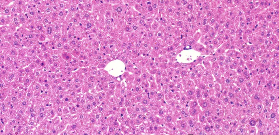 |
Mouse Liver Section (Hematoxylin and Eosin / H&E Stain)
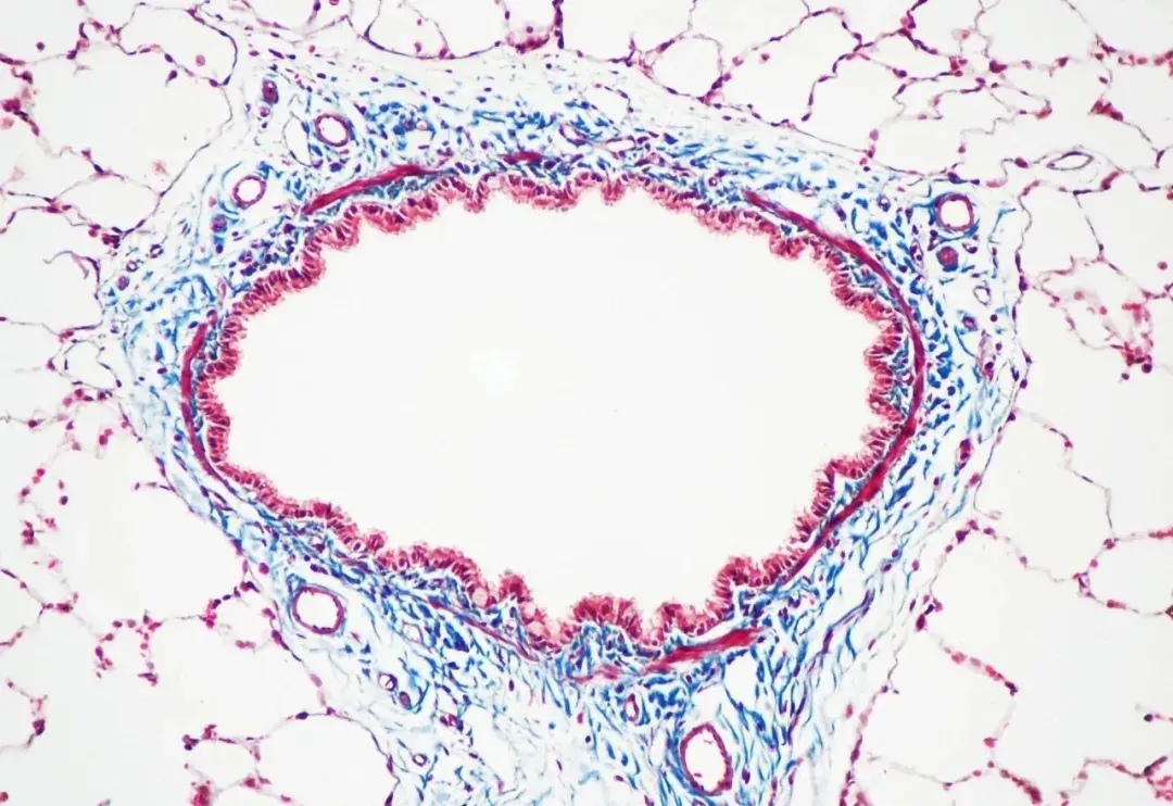 |
Rat Trachea Section (Masson’s Trichrome Stain: Connective tissue stained blue, cell nuclei stained purple, cytoplasm stained pink)
 |
Vascular Leiomyoma Section (Van Gieson Stain: Smooth muscle fibers stained yellow, collagen fibers stained red)
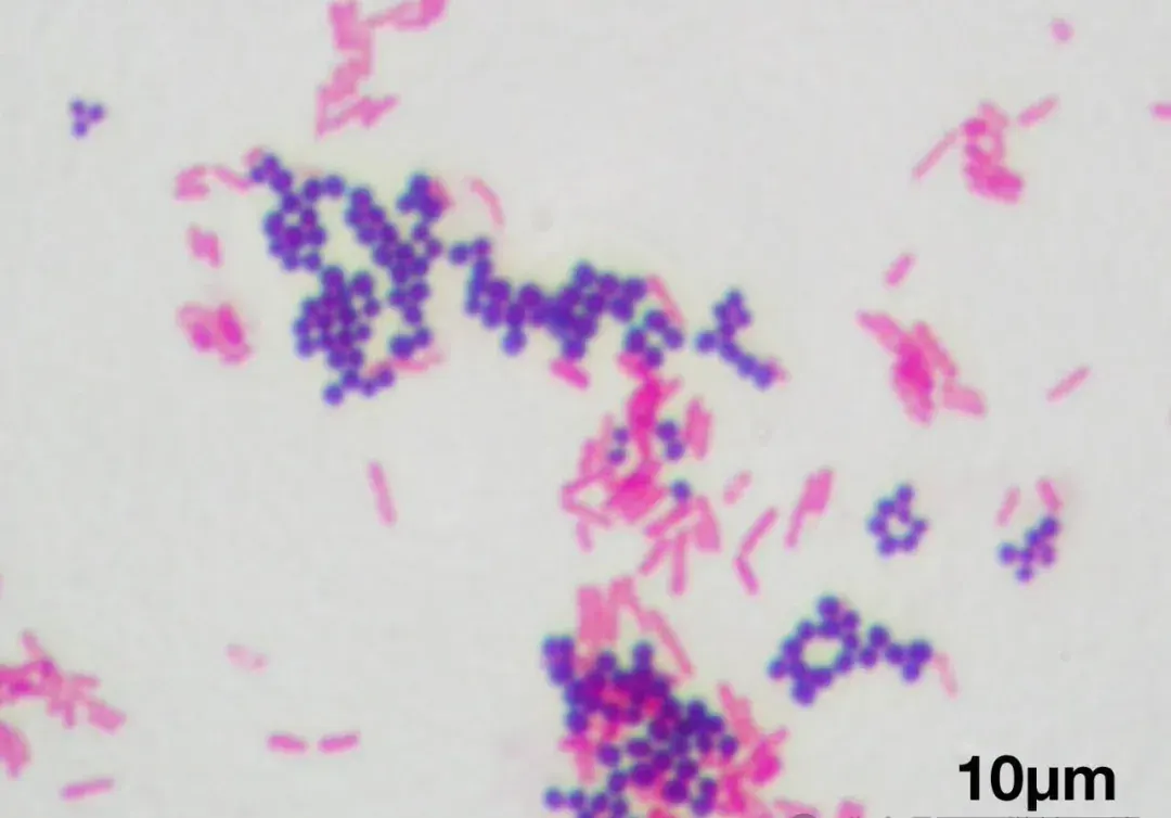 |
Gram-Stained Bacterial Smear (Gram-positive Staphylococcus aureus strain ATCC 25923 in purple; Gram-negative Escherichia coli strain ATCC 11775 in pink)
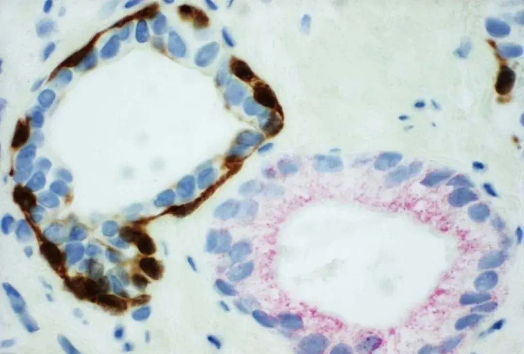 |
Immunohistochemical (IHC) Staining (PIN-4 staining of benign prostate glands on the left versus prostatic adenocarcinoma on the right, adenocarcinoma cells appear red)
02 | Darkfield Microscopy
Imaging Principles
Darkfield microscopy uses oblique illumination to block direct light from entering the objective lens.
Only light scattered by the sample enters the objective lens.
The background appears black while the specimen highlights brightly.
This method collects scattered light rays exclusively.
Primary Applications
- Live, unstained bacteria.
- Colloidal suspensions and nanoparticles.
- Small transparent structures and cellular appendages.
Key Image Characteristics
The high-contrast image features a dark background with brilliant sample illumination.
It increases visibility for thin, transparent specimens without staining.
The system is sensitive to specimen edges and minute particles.
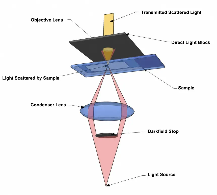
Darkfield Microscope Light Path Configuration
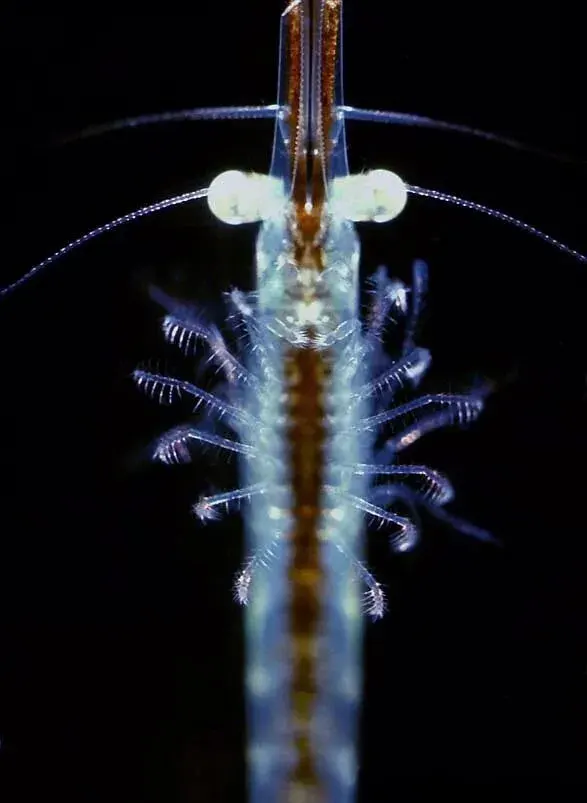
Darkfield Microscope Image showing dark background contrast
Typical Darkfield Examples
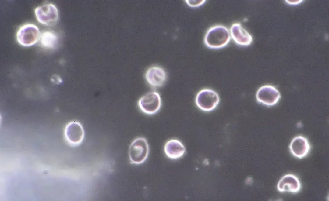
Unstained Erythrocytes (Red Blood Cells) under Darkfield Illumination
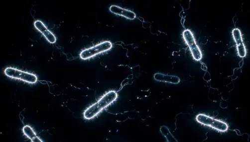
Live Bacterial Suspensions
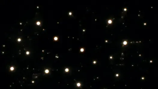
Colloidal Particles in Liquid Suspension
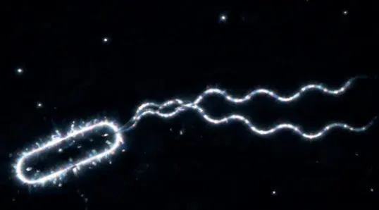
Bacterial Flagella and Fine Surface Structures
03 | Phase Contrast Microscopy
Imaging Principles
Phase contrast microscopy converts phase shifts from transparent samples into amplitude differences.
Small phase shifts$ Δϕ = \frac{2π}{λ} · (n₂ – n₁) · d $occur as light travels through biological structures with varying refractive indices.
The ring-shaped phase plate shifts light phases by$ \frac{π}{2} $ to generate destructive interference.
This optical transformation renders invisible cellular structures clearly visible as intensity changes.
Primary Applications
- Living, unstained cell cultures.
- Cellular growth and migration monitoring.
- Unstained biological specimens and fluid smears.
Key Image Characteristics
Researchers observe living cells in real time without fixatives or stains.
It remains a standard inspection tool in life science laboratories.
Images display characteristic halo artifacts around thick specimen boundaries.
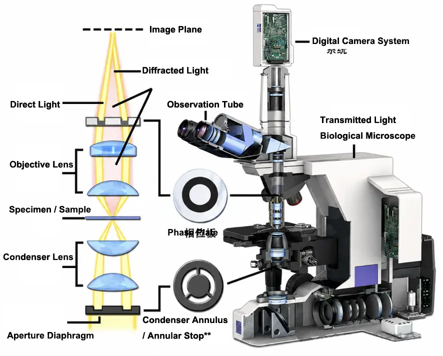
Phase Contrast Optical Path Diagram (Annular Diaphragm & Phase Plate)
Typical Phase Contrast Examples
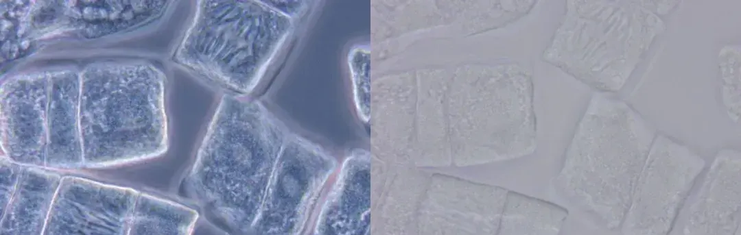
Unstained Onion Root Tip Cell Smear (Left: Leica HiPlan 100x/1.25 oil Ph3 objective with Ph3 annulus illumination; Right: Standard Brightfield illumination)
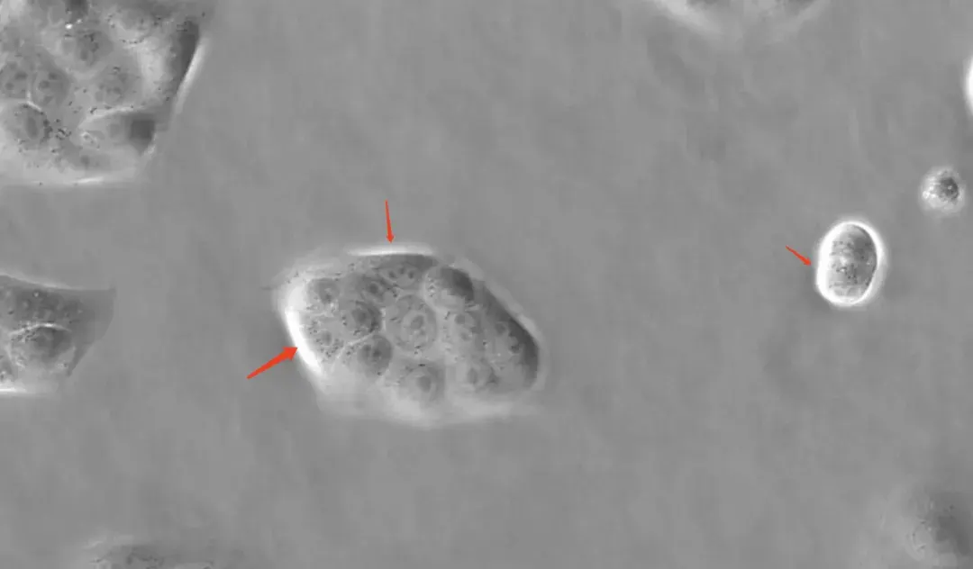
Madin-Darby Canine Kidney (MDCK) Cells (Red arrows mark bright halo optical artifacts)