 |
1. Introduction: The Cornerstone of Material Characterization
In the intricate landscape of modern materials science and industrial quality assurance, the metallurgical microscope (View HINOTEK metallurgical microscope Category) serves as the fundamental instrument for microstructural analysis. Unlike biological microscopy, which predominantly deals with transparent specimens and transmitted light, metallurgical microscopy is the science of the opaque. It is the optical gateway to understanding the internal architecture of metals, ceramics, composites, and semiconductors—materials that define the physical infrastructure of the modern world. For HINOTEK, standing at the intersection of global instrument supply and technical expertise, the metallurgical microscope is not merely a product category but a critical analytical tool that ensures the safety of aerospace components, the reliability of automotive drivetrains, and the performance of microelectronic circuits.
The necessity for such instruments arises from the physical nature of engineering materials. Metals and alloys absorb visible light within a few nanometers of their surface. Therefore, the traditional diascopic (transmitted) illumination used in biology is rendered useless. Instead, metallurgical microscopes employ episcopic (reflected) illumination, a sophisticated optical arrangement where the objective lens simultaneously acts as the condenser, projecting light onto the specimen and retrieving the image-forming rays reflected from the surface. This coaxial illumination path distinguishes the metallurgical microscope from all other optical instruments, dictating its mechanical design, optical coatings, and operational protocols.
This comprehensive report provides an exhaustive technical analysis of the metallurgical microscope. It dissects the physics of reflected light optics, the mechanical architecture of upright and inverted systems, the chemistry of metallographic sample preparation, and the standardized methodologies (ASTM/ISO) used in quantitative analysis. It is designed to serve as a foundational reference for laboratory managers, metallurgists, and quality control engineers navigating the HINOTEK platform for instrumentation solutions.
2. Physics of the Optical System
The performance of any optical microscope is governed by the laws of wave optics. However, in metallurgical systems, the behavior of light is complicated by the reflective nature of the specimen and the requirement for the illumination to traverse the objective lens twice. Understanding these physical principles is essential for maximizing resolution, contrast, and image fidelity.
2.1 The Vertical Illuminator and Beam Path
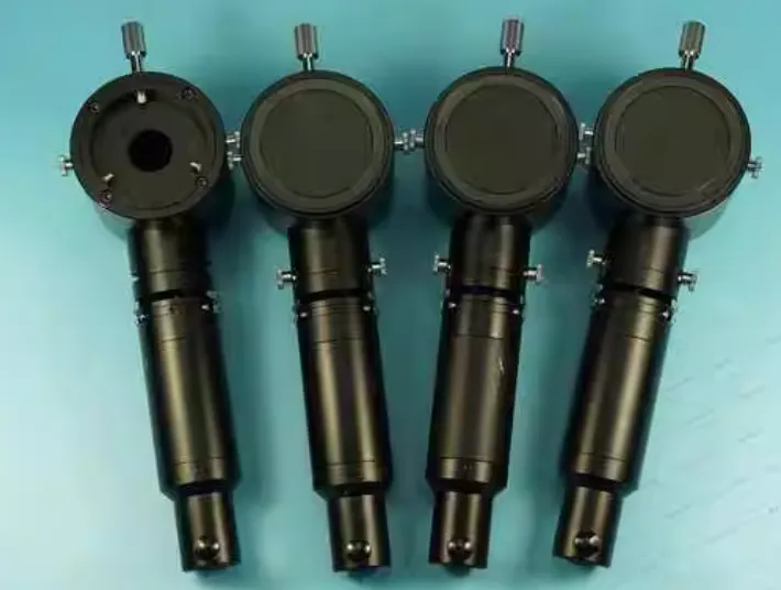 |
The vertical illuminator, often referred to as the epi-illuminator, is the heart of the metallurgical microscope. It is positioned between the observation tube (eyepieces) and the nosepiece (objectives). Its primary function is to inject illumination into the optical axis while simultaneously allowing the image-forming rays to pass through to the observer or camera sensor.
The optical path in a HINOTEK-standard metallurgical microscope typically follows a complex sequence designed to minimize internal reflections (flare) and maximize signal-to-noise ratio:
- Light Source Generation: Illumination begins at the lamp housing, typically housing a Halogen bulb or a high-intensity LED. The light is collected by a collector lens, which projects the filament image into the optical train.
- Aperture and Field Diaphragms: The light passes through two critical iris diaphragms. The Field Diaphragm controls the area of the specimen that is illuminated, serving to reduce stray light and glare from outside the field of view. The Aperture Diaphragm controls the angle of the cone of light entering the objective, directly influencing the numerical aperture (NA) of the illumination, which dictates resolution, contrast, and depth of field.
- The Beam Splitter: This is the critical junction. A semi-transparent mirror or prism (often with a 50/50 transmission/reflection ratio) reflects the horizontal light beam 90 degrees downward into the objective lens.
- The Objective as Condenser: The light travels down through the objective lens. In this phase, the objective functions as a condenser, focusing the light onto the specimen surface. This requires the objective to be corrected for glare, as high-intensity light is passing through the glass elements towards the sample.
- Specimen Interaction: The light strikes the opaque specimen. Rays hitting flat, perpendicular surfaces (like a polished grain interior) are reflected back up into the objective. Rays hitting sloped surfaces or etched grain boundaries are scattered away from the optical axis.
- Image Formation: The reflected rays re-enter the objective lens, which now acts as the imaging lens. They travel back up to the beam splitter, where they are transmitted (passed through) to the tube lens and eyepieces.
2.2 Numerical Aperture (NA) and Resolution Limitations
The resolving power of a metallurgical microscope—its ability to distinguish two closely spaced points as separate entities—is not infinite. It is strictly limited by the diffraction of light. The primary determinant of resolution is not magnification, but the Numerical Aperture (NA) of the objective lens.
The NA is a dimensionless number that characterizes the range of angles over which the system can accept or emit light. It is defined by the equation:
$$\text{NA} = n \cdot \sin(\mu)$$
Where:
- $n$ is the refractive index of the medium between the objective front lens and the specimen. For dry metallurgical objectives, this is air ($n \approx 1.0$). For immersion objectives, it is oil ($n \approx 1.51$).
- $\mu$ is the half-angle of the maximum cone of light that can enter the objective.
According to the Rayleigh criterion, the minimum resolvable distance ($r$) is calculated as:
$$r = \frac{0.61 \lambda}{\text{NA}}$$
Where $\lambda$ is the wavelength of the illumination light (typically 550 nm for green light). This relationship dictates that to resolve finer details (smaller $r$), one must either decrease the wavelength (using blue filters or UV light) or increase the NA.
For a standard high-quality dry metallurgical objective (100x magnification) with an NA of 0.95, the theoretical resolution limit is:
$$r = \frac{0.61 \times 550 \text{ nm}}{0.95} \approx 353 \text{ nm} \approx 0.35 \mu\text{m}$$
This physical limit implies that features closer together than 0.35 micrometers cannot be resolved as distinct separate points, appearing instead as a single blurred entity. This limitation is critical when analyzing fine lamellar structures in pearlite or nano-scale precipitates in superalloys.
2.3 Depth of Field (DOF) in Reflected Light
In metallography, samples are ideally polished flat, making depth of field less critical than in biological applications involving thick tissue sections. However, when examining fracture surfaces (fractography) or etched samples with high relief, depth of field becomes a limiting factor.
The depth of field correlates inversely with the square of the NA, making high-resolution imaging notoriously sensitive to focus. The formula for DOF is often approximated as:
$$\text{DOF} = \frac{\lambda}{n \cdot \text{NA}^2} + \frac{n}{M \cdot \text{NA}} \cdot e$$
Where:
- $M$ is the magnification.
- $e$ is the smallest distance the human eye can resolve (visual acuity limit, typically 0.25 mm projected onto the virtual image).
For a 100x objective with NA 0.90, the depth of field is less than 1 micrometer. This necessitates precise fine-focus mechanisms on HINOTEK instruments, often utilizing coaxial gear systems capable of micron-level adjustments. It also highlights the importance of sample flatness; a slightly tilted sample will have only a narrow band in focus at high magnification.
2.4 Optical Aberrations and Corrections
Metallurgical objectives must be highly corrected to produce accurate images. The “Plan” designation found on HINOTEK instrument specifications refers to Flat Field correction. A standard lens naturally projects a curved image (Petzval curvature), where the center is in focus but the edges are blurry. “Plan” objectives utilize additional lens elements to flatten this field, which is essential for photomicrography and digital image analysis where the entire sensor area must be in focus.
Furthermore, color correction is vital.
- Achromatic: Corrects for two colors (Red, Blue) and spherical aberration for one (Green).
- Apochromatic (Apo): Corrects for three or four colors chromatically and spherical aberration for two colors. These objectives, often containing fluorite elements, offer the highest performance and true color reproduction, crucial for identifying colored phases like copper inclusions or titanium nitrides.
3. Mechanical Architecture and Configuration
The mechanical design of a metallurgical microscope is dictated by the size and nature of the samples it is intended to examine. HINOTEK offers two primary configurations: Upright and Inverted.
3.1 Upright Metallurgical Microscopes
In the upright configuration, the optical train is positioned above the stage, looking down at the sample. This layout is similar to a standard biological microscope but with key differences in the stage and illumination.
- Sample Constraints: The sample is placed on top of the stage. Because the objective lens is fixed relative to the optical axis, the sample surface must be perfectly perpendicular to the beam. This requires the sample to be leveled. Usually, samples are mounted in resin pucks (phenolic or epoxy) with parallel faces, or placed on a bed of clay using a leveling press.
- Advantages: Upright microscopes often provide superior ergonomics for the operator during extended viewing sessions. They allow for easy observation of the sample surface orientation and are generally preferred for research applications where samples are small and carefully prepared.
- Limitations: The working distance (vertical space between stage and objective) is limited. Large industrial parts, such as a crankshaft or a large casting, cannot fit under the objectives.
3.2 Inverted Metallurgical Microscopes
The inverted microscope turns the optical arrangement upside down. The objectives are positioned below the stage, pointing upward, while the illuminator and viewing head remain above or to the side. The stage has an aperture (a central hole) through which the objective “sees” the sample.
- Operational Efficiency: The sample is placed face-down on the stage. The polished surface naturally aligns with the stage plane, eliminating the need for leveling. Gravity ensures the surface of interest is flat against the stage plate.
- Large Sample Capacity: Because the bulk of the sample extends upward, away from the objectives, there is virtually no limit to the height of the sample. Heavy industrial parts, cross-sections of pipes, or large automotive components can be placed directly on the stage.
- Advantages: This configuration is the workhorse of industrial quality control (QC) laboratories and foundries. It offers higher throughput because the leveling step is eliminated.
- Risk Factors: The objectives are vulnerable to gravity. Dust, grinding debris, or fluids (etchants/immersion oil) can fall from the sample surface down onto the objective front lens. HINOTEK inverted systems typically employ sealed nosepieces or protective glass slides to mitigate this risk.
3.3 Stage Mechanics and Specimen Handling
The mechanical stage of a metallurgical microscope must be significantly more robust than that of a biological scope. While a biological slide weighs a few grams, a mounted steel specimen or an unmounted casting can weigh several kilograms.
- Heavy-Duty Linkage: HINOTEK metallurgical stages often utilize steel-on-steel rack and pinion gears or heavy-duty wire drives to prevent “stage creep” (unwanted downward drift due to weight).
- Surface Inserts: Inverted microscope stages come with various metal inserts (teardrop, circle, rectangular) with different aperture sizes. These prevent small mounted samples from falling through to the objectives while supporting large irregular parts.
- Coaxial Controls: Precise X-Y movement is controlled by low-position coaxial knobs, allowing the operator to scan the sample surface systematically—a requirement for standardized inclusion rating or grain size surveys.
4. Imaging Modes and Contrast Techniques
While Brightfield is the standard imaging mode, the complex nature of metallic microstructures often requires advanced contrast techniques to reveal features that are invisible under normal illumination.
4.1 Brightfield (BF) Illumination
In Brightfield, the light impinges on the specimen surface perpendicular to the plane of the stage.
- Mechanism: Light travels through the objective, hits the sample, and reflects back.
- Contrast Generation: Flat, smooth areas (like the polished face of a ferrite grain) reflect light directly back into the objective and appear bright. Rough features, such as etched grain boundaries, cracks, or pits, scatter light away from the objective aperture and appear dark.
- Application: This is the default mode for grain size estimation, porosity measurement, and general microstructural characterization.
4.2 Darkfield (DF) Illumination
Darkfield illumination is a technique used to enhance the visibility of edges, scratches, and grain boundaries that might be too subtle for Brightfield. It requires specialized “BD” (Brightfield/Darkfield) objectives.
- Objective Construction: BD objectives are larger than standard ones because they contain a hollow collar surrounding the central lens elements.
- Optical Path: In the vertical illuminator, a central stop blocks the middle of the light beam, creating a hollow cylinder of light. This light travels down the outside collar of the objective. A ring mirror or prism at the tip of the objective directs this light inward at a steep oblique angle toward the sample.
- Contrast Mechanism: If the sample surface is perfectly flat, the oblique light reflects off the surface at the same angle and misses the central objective lens entirely. The background appears pitch black. However, if there is a scratch, edge, or particle, it scatters the light in all directions. Some of this scattered light enters the central lens.
- Result: Defects glow bright white against a dark background. This is critical for semiconductor wafer inspection (detecting dust or scratches) and for identifying fine grain boundary precipitates.
4.3 Differential Interference Contrast (DIC)
Also known as Nomarski contrast, DIC is a powerful technique for visualizing surface topography (slope) that is too shallow to generate contrast in Brightfield. It creates a pseudo-3D relief image.
- Optical Components: DIC requires a polarizer, an analyzer, and a specialized Nomarski (or Wollaston) prism located above the objective.
- Physics of Shear: The prism splits a linearly polarized light beam into two orthogonally polarized rays (Ordinary and Extraordinary). These rays are spatially offset (sheared) by a distance slightly smaller than the resolution of the objective.
- Phase Shift: The two rays hit the sample at adjacent points. If there is a microscopic slope (height difference) between these two points, one ray travels a slightly different path length than the other.
- Interference: The rays reflect, travel back through the prism where they are recombined, and pass through the analyzer. The analyzer causes the two rays to interfere. The phase difference caused by the slope is converted into an amplitude (brightness) difference.
- Visual Result: One side of a slope appears bright, and the other appears dark, creating a relief effect. This allows metallographers to see slip bands, twinning, and hardness gradients on polished, unetched surfaces.
4.4 Polarized Light Microscopy
This mode utilizes a polarizer (in the illumination path) and an analyzer (in the viewing path) crossed at 90 degrees.
- Isotropic Materials: Materials with a cubic crystal structure (like steel, aluminum, copper) reflect light without changing its polarization state. Under crossed polars, they appear dark (extinction).
- Anisotropic Materials: Materials with non-cubic structures (like magnesium, zinc, titanium, or graphite) are birefringent. They rotate the plane of polarization of the incident light. When this rotated light hits the analyzer, a component passes through, causing the grain to appear bright or colored.
- Applications:
- Cast Iron: Verifying the nodularity of graphite. Graphite nodules exhibit a characteristic “Maltese Cross” interference pattern under polarized light.
- Titanium/Zirconium: Grain size analysis is often performed under polarized light because these metals are difficult to etch chemically but show distinct grain coloration in polarized light (especially when anodized).
5. The Science of Metallographic Sample Preparation
A metallurgical microscope, no matter how advanced, can only image the surface presented to it. If the sample preparation is poor—filled with scratches, deformation, or staining—the resulting image will be misleading. The goal of metallography is to reveal the true microstructure, not artifacts of the preparation process.
5.1 Sectioning
The first step involves cutting the component to a size suitable for mounting.
- Abrasive Cutters: These use rotating wheels (Silicon Carbide or Alumina) flooded with coolant. The critical parameter here is heat control. Excessive heat during cutting can alter the microstructure (e.g., “burning” steel can create false martensite layers).
- Precision Saws: For delicate electronics or brittle ceramics, low-speed diamond wafering saws are used to minimize mechanical damage.
5.2 Mounting
Mounting encapsulates the sample in a polymer to provide a safe handle and to protect the edges of the sample (which are often the most critical areas).
- Compression Mounting (Hot Press): The sample is placed in a cylinder, covered with resin powder (Phenolic/Bakelite or Epoxy), and subjected to heat ($150^\circ$C) and pressure (20-30 MPa). This produces the hardest, most durable mount with the best edge retention.
- Castable Mounting (Cold Mount): For heat-sensitive materials (like PCBs with low-melting solders), a two-part liquid resin (epoxy or acrylic) is poured over the sample and allowed to cure at room temperature.
5.3 Grinding and Polishing
This process involves sequential material removal to create a scratch-free surface. It is typically divided into Planar Grinding, Fine Grinding, and Polishing.
Table 1: Standard Grinding/Polishing Recipe for Carbon Steel (e.g., 1045)
| Step | Abrasive Media | Lubricant | Force (N) | Speed (RPM) | Time | Purpose |
| Planar Grinding | SiC Paper 240 Grit | Water | 25 | 300 | Until Flat | Remove sectioning damage and level surface. |
| Fine Grinding | SiC Paper 400 Grit | Water | 25 | 150 | 1 min | Refine surface scratches. |
| Fine Grinding | SiC Paper 600 Grit | Water | 25 | 150 | 1 min | Further refinement. |
| Fine Grinding | SiC Paper 800/1000 Grit | Water | 25 | 150 | 1 min | Pre-polish finish. |
| Rough Polish | Diamond Suspension 9 $\mu$m | Extender | 20 | 150 | 3 min | Remove grinding deformation. Use hard cloth. |
| Intermediate | Diamond Suspension 3 $\mu$m | Extender | 20 | 150 | 3 min | Remove micron-level scratches. |
| Final Polish | Diamond Suspension 1 $\mu$m | Extender | 15 | 150 | 2 min | Optical mirror finish. |
| Chemo-Mechanical | Colloidal Silica (0.05 $\mu$m) | Water | 10 | 150 | 1 min | Removes final “smear” layer and deformation. |
Special Considerations for Aluminum (e.g., 6061 Alloy):
Aluminum is soft and susceptible to embedding abrasive particles. Water can sometimes cause corrosion or staining.
- Lubrication: Often requires paraffin-based lubricants instead of pure water.
- Final Polish: A long final polish with Colloidal Silica is crucial to remove the “smear layer” (a layer of flowed metal that hides the true grain structure).
5.4 Etching Chemistry
A polished metal surface usually appears as a featureless mirror. To see the grains, we must chemically attack the surface.
- Grain Boundary Attack: Grain boundaries are regions of high energy due to atomic mismatch. When exposed to an etchant, these boundaries dissolve faster than the grain interiors, creating tiny grooves. Under the microscope, these grooves scatter light and appear as dark lines.
- Common Etchants:
- Nital (2-5% Nitric Acid in Ethanol): The standard for carbon steels. Reveals ferrite grain boundaries and pearlite lamellae.
- Keller’s Reagent: Containing HF, HCl, and $HNO_3$. Standard for Aluminum alloys.
- Kroll’s Reagent: For Titanium alloys.
- Picral: Picric acid in ethanol. Excellent for revealing fine carbides in heat-treated steels without etching the ferrite grain boundaries too deeply.
6. Quantitative Metallography and Standards
Metallurgical microscopy is a quantitative science. HINOTEK instruments are often the source of data for certification according to international standards (ASTM, ISO, JIS).
6.1 Grain Size Analysis (ASTM E112)
Grain size is a fundamental property that dictates mechanical behavior. Fine grains generally lead to higher strength and toughness (Hall-Petch relation). ASTM E112 is the primary standard for measuring this.
Methods:
- Comparison Method: The operator views the sample and visually compares it to a standard wall chart or an eyepiece reticle containing reference hexagons. This is fast but subjective.
- Intercept Method (Heyn Procedure): A grid of test lines is overlaid on the image. The operator (or software) counts the number of times grain boundaries intercept these lines. The ASTM Grain Size Number ($G$) is calculated as:
$$G = (6.643856 \log_{10} \bar{N}_L) – 3.288$$
Where $\bar{N}_L$ is the number of intercepts per unit length (mm). This method provides a statistically rigorous value with defined confidence intervals.
6.2 Inclusion Rating (ASTM E45)
Non-metallic inclusions (oxides, sulfides, silicates) are inevitable byproducts of steelmaking. They act as stress concentration points and can initiate fatigue cracks. ASTM E45 prescribes the method for rating the “cleanliness” of steel.
Inclusion Types:
- Type A (Sulfides): Elongated, gray stringers. Malleable.
- Type B (Aluminates): Fragmented, black rows. Brittle.
- Type C (Silicates): Elongated, black/gray. Malleable but often wider than sulfides.
- Type D (Globular Oxides): Spherical, scattered spots. Non-deformable.
Rating Process:
The sample is scanned (typically 160 $mm^2$ area). The “Worst Field” method (Method A) involves finding the field of view with the most severe inclusions and comparing it to a standard chart (JK Chart). Modern automated HINOTEK systems use Method D, where an image analyzer scans the entire sample and calculates histograms of inclusion length and thickness, providing a far more accurate assessment of steel quality.
7. Industrial Applications and Case Studies
7.1 Automotive and Aerospace: Failure Analysis
In the automotive industry, metallurgical microscopes are the first line of defense in failure analysis.
- Case Study: A broken transmission gear. The metallographer examines the fracture surface.
- Observation: Presence of “beach marks” indicates fatigue failure.
- Microstructure: A cross-section reveals that the carburized case (hardened outer layer) was too thin, or that there was “intergranular oxidation” at the surface, weakening the grain boundaries.
- Conclusion: The heat treatment process atmosphere was incorrect.
7.2 Electronics: PCB Cross-Sectioning
Printed Circuit Boards (PCBs) are complex composites of copper, fiberglass (FR4), and solder.
- Application: “Micro-sectioning” coupons are cut from the PCB panel.
- Target: The plated through-hole (via). The microscope measures the thickness of the copper plating on the hole wall (typically 25 microns required).
- Defect Detection: Inspectors look for “knee cracks” (cracks at the corner of the via), “delamination” (separation of fiberglass layers), or “voids” in the solder joints. Darkfield illumination is particularly useful here to make delaminations “glow”.
7.3 Foundries: Cast Iron Nodularity
In ductile cast iron, the graphite must form spheres (nodules) to provide ductility. If the magnesium treatment fails, the graphite forms flakes (gray iron), which is brittle.
- Analysis: Using a metallurgical microscope (often with image analysis software), the “Nodularity %” is calculated.
- Criteria: >90% nodularity is often required for safety-critical parts like suspension arms. This analysis is usually performed on “as-polished” (unetched) samples to maximize the contrast between the black graphite and the bright metal matrix.
8. Maintenance and Troubleshooting
A metallurgical microscope is a precision optical instrument. Its performance degrades rapidly if not maintained.
8.1 Optical Cleaning Protocols
Dust and oil are the primary enemies.
- Dry Objectives: These lenses are designed for use in air. If they accidentally touch immersion oil, the oil can seep into the spring-loaded nose and dissolve the cement holding the lens elements. Immediate cleaning with lens tissue and a solvent (like an ether-alcohol mix or commercial lens cleaner) is required.
- Oil Immersion Objectives: Must be cleaned after every session. Dried immersion oil turns into a hard, sticky resin that is extremely difficult to remove without damaging the anti-reflective coatings.
8.2 Troubleshooting Image Quality
Table 2: Common Metallurgical Microscope Issues and Solutions
| Symptom | Probable Cause | Corrective Action |
| Blurry / Hazy Image | Dirty Objective Front Lens | Clean lens carefully with solvent and lens tissue. |
| “Hot Spot” in center | Field Diaphragm too open | Close Field Diaphragm until it frames the field of view. |
| Low Contrast / Washed out | Aperture Diaphragm too open | Close Aperture Diaphragm to 70-80% of the Objective NA. |
| One side out of focus | Sample not level | Re-mount sample or use leveling press/clay. |
| Dark Corners (Vignetting) | Nosepiece not centered | Rotate nosepiece until it “clicks” firmly into detent. |
| Stage drifts downward | Tension loose | Adjust coarse focus tension ring. |
9. The Digital Frontier
The integration of digital cameras and software has transformed metallurgical microscopy from a subjective art to an objective data science.
- Large Area Tiling (Stitching): HINOTEK systems now support automated stages that scan a large sample (e.g., 50mm x 50mm) at high magnification. The software stitches thousands of individual images into a single “gigapixel” map, allowing users to zoom from the macro view of the whole part down to the micro view of a single grain boundary.
- Extended Depth of Focus (EDF): By capturing a “Z-stack” of images at different focal heights, software can extract the sharpest pixels from each layer and combine them. This creates a fully focused image of a rough fracture surface that would otherwise be impossible to image with a standard optical microscope.
Conclusion
The metallurgical microscope stands as a pillar of industrial science. It bridges the gap between the macroscopic world of engineering specifications and the microscopic world of atomic arrangements. For HINOTEK, providing these instruments involves more than mere supply; it requires a commitment to understanding the rigorous demands of sample preparation, the physics of optical contrast, and the precision of standardized analysis.
Whether determining the safety of a structural steel beam, the conductivity of a microchip, or the longevity of an engine component, the metallurgical microscope provides the visual and quantitative data upon which modern industry relies. Through the selection of appropriate optics, robust mechanical platforms, and advanced imaging techniques, laboratories can ensure that the materials shaping our future meet the highest standards of quality and reliability.
Works cited
- Understanding Metallurgical Microscope – Complete Guide for Industrial and Research Applications
- How Does A Metallurgical Microscope Work? – Prime Lab Med
- Introduction to Reflected Light Microscopy – Evident Scientific
- Microscopy Basics | Illumination and the Optical Train – Zeiss Campus
- Optical/ Metallurgical Microscopy | PPTX – Slideshare
- Reflected Light Microscopy – Zeiss Campus